- Published: 2025-09-08
- Updated: 2025-09-09
- MeshLib Team
What is a CT Scan with 3D Reconstruction?
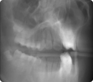
CT 3D reconstruction is the process of converting a series of 2D CT scans captured from different angles into 3D volumes. In practice, it suggests combining multiple X-ray images taken from a full 360-degree rotation (the more scans, the better the quality) to generate a detailed 3D representation of the target area of interest. This provides for sharper diagnoses, precise surgical planning, treatment monitoring, radiology, and patient-specific 3D printing.
How to do 3D Reconstruction of CT Image Files?
CT 3D Reconstruction starts right when projection data leaves the scanner. Speaking of modern toolkits, they group their algorithms into two broad families:
- Analytic solvers convert projections to voxels in a single pass. They excel at speed, trimming overall 3D reconstruction CT scan time to minutes. However, they amplify noise if the scan was acquired at a low dose.
- Iterative solvers refine the volume through multiple forward-projection cycles. Each round cross-checks the synthetic image against the real detector data, driving down streaks, rings, and beam-hardening artifacts.
Anyway, proper noise reduction and data processing are critical. Even a minor error on every slice can snowball into millimetre-scale deviations across the stack. This is unacceptable for surgical planning or printed implants. Best-practice pipelines, therefore:
- Run adaptive, edge-preserving filters before reconstruction;
- Store interim voxel tiles in a fast database cache;
- Apply artifact detectors during each iteration so problems are fixed early.
Delving into what we could offer you in terms of CT 3D reconstruction: our library starts by ingesting a stack of radiograph images and running our CT Reconstruction algorithm. The latter obtains dense volumes of voxel data from a number of radiograph images. Once the voxel grid is in memory, users can open a Volume Rendering view for instant visual checks and employ Iso-value Adjustment to isolate a target tissue or material density on the go. In this fashion, large CT datasets turn into analyzable 3D volumes quickly while preserving resolution.
After CT 3D reconstruction, our Voxel-to-Mesh Conversion routines can help you create a surface model. The latter can be processed through a Mesh Healing round to detects and fix mesh issues and make sure that your output is error-free (however, in most cases, voxel-based meshes are of good quality).
How to Do Computed Tomography 3D Reconstruction in MeshLib?
CT 3D reconstruction converts raw CT projection data into a volumetric dataset of density voxels by applying Filtered Back Projection (FBP). This voxel volume can then be used directly for interactive 3D visualization, segmentation, or rendering. In the DICOM workflow, the same volume may be stored as a stack of aligned 2D slices for compatibility and further processing.
Applications
In case you work in one of the following domains and need potent CT 3D reconstruction capabilities, we invite you to try MeshLib for these purposes:
- Medical imaging via CT scanners. Here, the desirable outcomes are attained by a combination of hardware (say, high-speed rotating gantries and advanced X-ray detectors) with iterative algorithms. By applying such CT methods, one gets clearer visuals of anatomical structures. In this fashion, more precise diagnoses and treatment planning become possible. That is to say, by reducing noise and enhancing resolution via different filters, CT reconstructions are instrumental in detecting and addressing a wide range of conditions.
- Industrial CT Scanners. First, industrial CT scanners capture internal data of complex components. CT 3D reconstruction algorithms then convert raw projections into a detailed volumetric model. This allows engineers to assess internal features without disassembling or damaging the part.
- Science, research, and academic activities. Well beyond immediate applications in industry or clinical settings, CT reconstruction also plays a pivotal role in teaching and exploratory research. Biomedical researchers develop and validate novel algorithms to improve image reconstruction quality or reduce motion/artifact effects in scanning. As for universities and research institutions, they employ specialized CT scanners in biomedical engineering, physics, and computer science programs to give students hands-on experience.
Step-by-Step Instructions
With MeshLib, the flow looks like this:
- In CT imaging, either the X-ray source and detector arrays rotate around the subject, or—when scanning small objects—the source and detector remain stationary while the object itself is rotated on a specialized turntable. In both cases, the purpose is to capture multiple 2D radiographic projections. It is important to note that these raw projections are not in DICOM format. They cannot be interpreted visually in their initial state. Instead, they must undergo a reconstruction process to transform them into slice-based image data.
- Then, our reconstruction algorithm processes the raw projection data to generate a volumetric dataset of density voxels using Filtered Back Projection. Once all projections have been incorporated, this 3D volume can be directly used for visualization, segmentation, or analysis. If needed, the volume may also be exported as a series of cross-sectional slices for compatibility.
The key capability of importance that you must be able to apply here:
- Filtered back projection, using mathematical filters to convert collected projection data into spatial domain information.
- Once reconstructed, our slices are often stored in the Digital Imaging and Communications in Medicine (aka DICOM) format. This is one of the most widely adopted standards for exporting, sharing, and archiving medical images.
DICOM not only encapsulates the image pixel data. It also preserves critical metadata, e.g., patient information, slice orientation, pixel spacing, etc. - After the volumetric dataset is assembled, post-processing techniques might be employed further:
- Volume rendering algorithms allow observers to view the entire 3D volume semi-transparently, revealing interior structures in a single visual representation;
- Segmentation involves partitioning the volumetric dataset into meaningful regions for precise measurements, 3D modeling, and computational analyses.
Video Overview
Performance
Among other thing, MeshLib is notable for the following advantages:
- On average, our algorithms, including the one for CT 3D reconstruction, can run up to 10× faster on average than other SDKs. Please note that CT reconstruction is currently supported only with CUDA. That is, this feature in MeshInspector, the solution based on MeshLib, is not available on macOS or in web-based environments;
- Our library opens scans in seconds, up to 5× faster than standard viewers, and flip through images smoothly, even with huge files.
Supported Programming Languages and Versions
- C++ core is fully cross-platform and runs anywhere with zero limitations.
- Python binding is supported on versions 3.8 through 3.13 across all major OSes:
- Windows: Python 3.8–3.13
- macOS: 3.8–3.13 (Intel builds start at 3.9)
- Linux: 3.8–3.13, packaged for any manylinux 2.31 or newer system
What our customers say
Thomas Tong
Founder, Polyga
Gal Cohen
CTO, customed.ai

Mariusz Hermansdorfer
Head of Computational Design at Henning Larsen Architechts

HeonJae Cho, DDS, MSD, PhD
Chief Executive Officer, 3DONS INC

Ruedger Rubbert
Chief Technology Officer, Brius Technologies Inc


Start Your Journey with MeshLib
MeshLib SDK offers multiple ways to dive in — from live technical demos to full application trials and hands-on SDK access. No complicated setups or hidden steps. Just the tools you need to start building smarter, faster, and better.